An axolotl can lose a leg to a hungry tankmate and regenerate a new one — bone, cartilage, muscle, nerve, skin, blood vessels, even the tiny pigment patterns — over several months. No scar. No prosthesis. No lingering deficit. The same animal, if you nick a section of its spinal cord or remove a piece of its heart ventricle, will rebuild those too, drawing on a biology that mammals appear to have walked away from somewhere deep in evolutionary time.
This is why labs in Vienna, Boston, Dresden, Wake Forest and Madison keep colonies of these pale Mexican salamanders alive in climate-controlled rooms, feeding them pellets and tracking every cell of every regrown limb under confocal microscopes. The axolotl is the closest thing biology has to a working blueprint for what humans used to be able to do, or might one day learn to do again.
The cell that knows how to start over
When an axolotl loses a limb, the wound seals within hours. A thin sheet of epidermal cells slides over the stump and thickens into something called the apical epithelial cap. Underneath, mature cells (muscle, cartilage, connective tissue) do something that mammalian cells almost never do. They dedifferentiate, reverting to a more plastic, progenitor-like state, and pile up into a mass called the blastema.
The blastema is the engine. It looks, under a microscope, almost identical to the limb bud of an embryo. It is, in effect, the animal reopening a developmental program it had closed decades earlier and running it again from a slightly altered starting condition.
That is the trick. Human wounds close with fibrosis. Collagen laid down fast and messily, producing scar. Axolotl wounds close with a pause, an inflammatory phase that clears debris without locking the tissue into a fibrotic dead end, and then a return to embryonic-style growth.
Vienna’s positional code
One of the long-standing puzzles was how the regrowing limb knows what to build. If you amputate at the shoulder, the axolotl grows a whole arm. If you amputate at the wrist, it grows a hand. The blastema cells somehow remember where on the body they sit.
In 2025, Elly Tanaka’s lab at the Institute of Molecular Biotechnology of the Austrian Academy of Sciences (IMBA) in Vienna, working out of the Vienna BioCenter alongside the Research Institute of Molecular Pathology, published a paper in Nature identifying a gene called Hand2 as a central piece of that positional code. Cells along the posterior side of the limb express Hand2 throughout life; cells on the anterior side do not. After amputation, those posterior cells dial Hand2 expression up, which switches on a Shh signal that radiates outward like a broadcast and tells the blastema which way is thumb and which way is pinky.
Disrupt Hand2, and the axolotl can no longer pattern a complete limb. Mutant animals regenerate with fewer digits than they started with, and in the most severe cases nothing forms beyond the shoulder girdle. Push the gene the other way. Express it on the anterior side, where it normally isn’t, and the same machinery overshoots in the opposite direction, producing extra digits or, occasionally, a second limb growing from the wrong place. The cellular hardware is intact in both cases. What changes is the map.
The holy-grail gene shared across species
The Vienna work answered the positional question. A separate collaboration, published in 2026 in the Proceedings of the National Academy of Sciences, asked a different one: is any of this conserved in animals that don’t regenerate well, including us?
Researchers at Wake Forest University and Duke University, comparing regenerating tissue across three species — axolotls, zebrafish, and mice — found that the epidermis of all three activated the same two genes, SP6 and SP8, during the early regenerative phase.
The conclusion was that universal genetic programs appear to drive regeneration across very different organisms, suggesting a deep evolutionary inheritance that mammals still carry but mostly leave silent.
When Josh Currie’s lab at Wake Forest used CRISPR to remove SP8 from the axolotl genome, the animals lost the ability to regenerate proper limb bones. The same was true in mice missing SP6 and SP8 in regenerating digit tips. And mice can regrow the very tips of their toes, a narrow capacity humans retain in fingertips when the nailbed survives.
David Brown’s lab at Duke then built a viral gene therapy delivering FGF8, the signaling molecule normally switched on by SP8. In treated mice, damaged digits showed renewed bone growth. It was partial, modest, and a long way from a regrown hand, but it was the first time the regenerative-epidermis program had been deliberately reignited in a mammal.
Why the heart matters
Limbs draw the cameras, but the heart is where the medical urgency lives. A human heart attack kills a patch of cardiac muscle that is replaced by scar tissue. The scar holds the organ together. It does not pump. Survivors live the rest of their lives with a weaker heart and a higher chance of failure.
An axolotl with a ventricular wound does not lay down a scar. Within weeks, cardiomyocytes near the injury re-enter the cell cycle, divide, and rebuild contractile tissue. The same is true of zebrafish, which can lose a substantial portion of their ventricle and regrow it over weeks. Both species use overlapping signaling cascades involving nerve-derived trophic factors, retinoic acid gradients, and the same kind of dedifferentiation programs that drive limb regrowth.
Mammalian hearts run a version of this program only in the first week of life. Newborn mice can regenerate ventricular tissue but lose this capacity during development. Something switches off. Finding that switch, and persuading it back on safely, is one of the live questions in cardiac regenerative medicine.
The spinal cord and the brain
Sever an axolotl spinal cord and the ependymal cells lining the central canal proliferate, migrate to the lesion, and rebuild the neural tube. Axons regrow across the gap. Motor function returns. The same animal can lose chunks of forebrain tissue and replace neurons over a period of weeks, with new cells integrating into existing circuits.
In mammals, a severed spinal cord forms a glial scar. A dense barrier of astrocytes that, paradoxically, both protects the surrounding tissue and blocks any attempt at axonal regrowth. The scar is the immune system doing its job in the only language it knows. The axolotl immune system, by contrast, runs an inflammatory phase that is shorter, more tightly regulated, and exits without leaving the fibrotic walls that define mammalian healing.
Single-cell RNA sequencing of axolotl blastemas has mapped this immune choreography in detail, identifying macrophage populations that appear to be required for regeneration. Deplete those macrophages, and the limb fails to regrow at all. The wound just sits there, open and stalled.
A genome built for this
The axolotl genome is enormous. About ten times the size of the human genome. Most of that bulk is repetitive elements and ancient retroelements expanded over evolutionary time, padding out the introns of otherwise familiar genes. For decades, sequencing it was considered nearly impossible. A chromosome-scale assembly finally arrived and revealed salamander-specific gene duplications that appear to function as regulatory switches during regeneration.
Comparative work has since shown that humans share most of the relevant protein-coding genes. What seems different is the regulatory wiring around them. When they switch on, in which cells, and for how long. The axolotl appears to have kept a developmental toolkit accessible into adulthood that mammals close down shortly after birth.
Why labs keep them alive
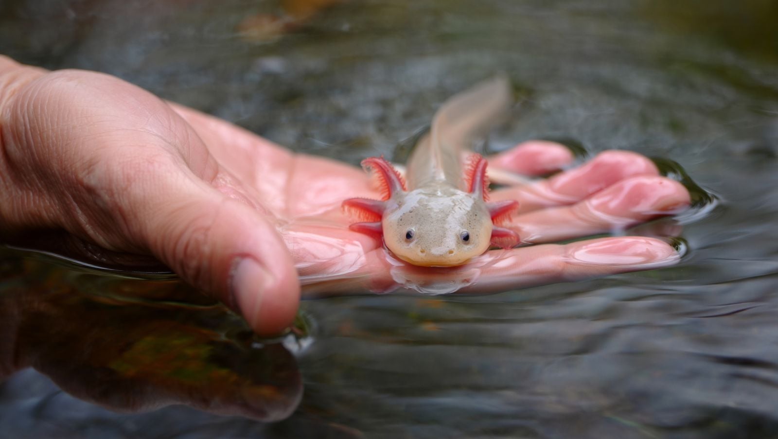
The Mexican axolotl is critically endangered in its native habitat, the canals of Xochimilco south of Mexico City. Pollution and invasive species have collapsed the wild population, and recent reintroduction efforts using captive-bred animals have only just begun to show survival in restored wetland refuges.
Yet there are more axolotls in laboratories worldwide than anywhere else on Earth. The Ambystoma Genetic Stock Center at the University of Kentucky maintains large breeding populations. Vienna, Dresden, Boston, Wake Forest, Madison, Hannover, and Tsukuba all maintain breeding colonies. Each animal lives in tank water cooled to around 18 degrees Celsius, fed earthworms or formulated pellets, photographed weekly, and tagged for genealogy.
They are kept alive because no engineered system, no stem cell scaffold, no bioprinted organ, no chemically induced pluripotency, has matched what they do casually, in a tank, while waiting for dinner.
The frontier of borrowed biology
The interest in axolotl biology sits inside a broader push to extend the human body’s repair window. Silicon Valley has been pouring money into cell reprogramming as an anti-aging frontier, betting that the Yamanaka factors and related techniques can roll mature cells back to a younger state. The axolotl does this naturally, in a controlled and localized way, every time it loses a body part.
Comparative genomics studies have made clear that the relevant pathways are not entirely missing from mammals. They are silenced, fragmented, or repurposed. The salamander has kept them assembled.
Whether human medicine can borrow the schematic remains an open question. The 2026 SP8 work in mice was a partial regrowth of bone in a digit tip, not a regrown arm. The Hand2 work in Vienna explained how positional information is stored in salamander cells, not how to install that storage in human ones.
The animal that doesn’t quite grow up
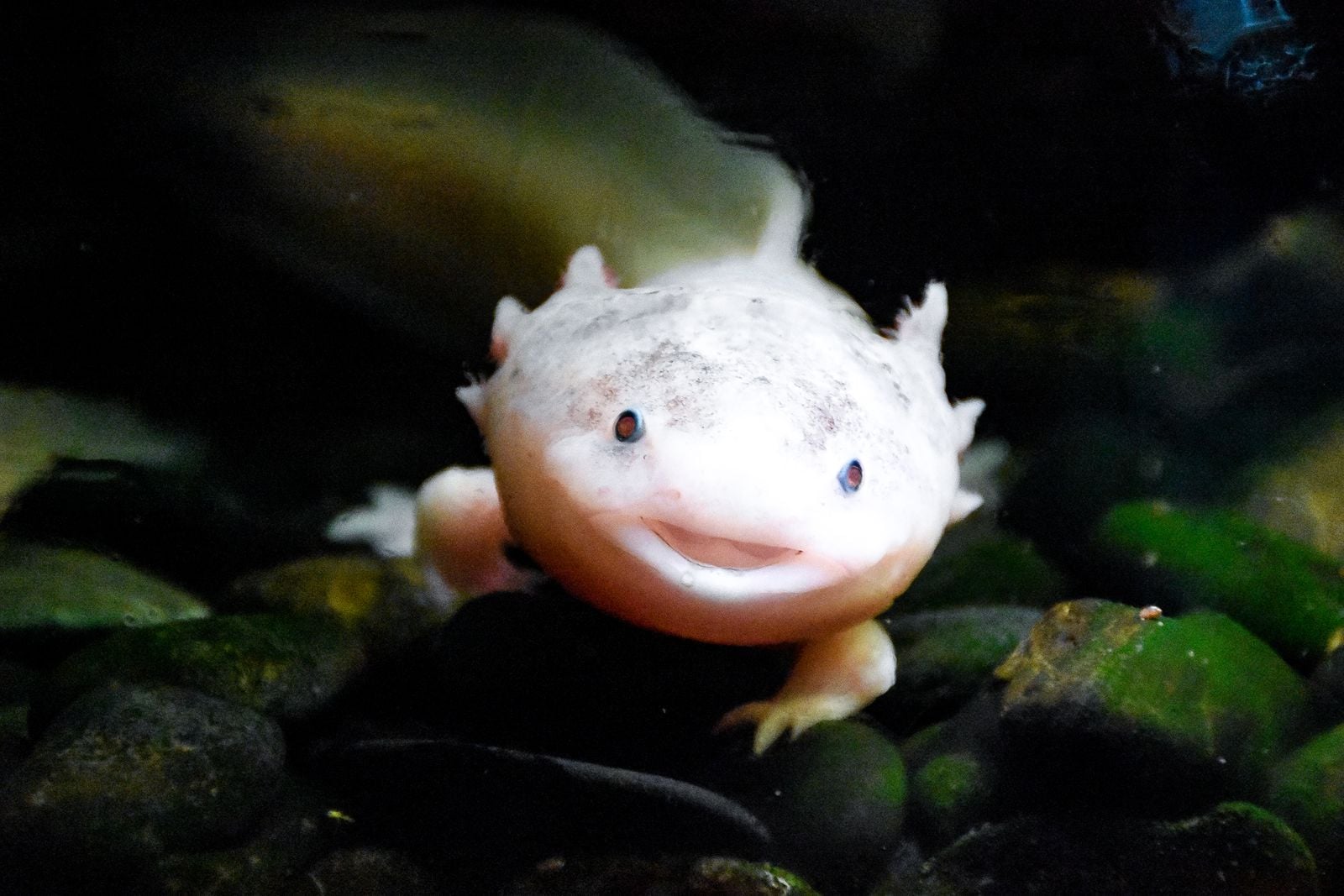
Axolotls keep their feathery external gills throughout adulthood, a trait called neoteny. They never undergo the metamorphosis that other salamanders go through. They reach sexual maturity while still in what looks, anatomically, like a larval form. It is tempting to draw a connection between this arrested development and the regenerative capacity, as if the animal stays close enough to its embryonic biology to keep the toolbox open.
An adult axolotl in a Vienna tank, missing a foreleg from a tankmate squabble three weeks earlier, will already have a translucent bud at the stump. Inside that bud, cells are doing something a human body has not done since the first month after conception. Bone is being patterned. Nerves are being routed. A thumb is being decided on.
The bud is roughly the size of a grain of rice. In four months, it will be a leg.